
This is how life begins - the journey from the moment we are born. From a single cell, we grow into the most complex creatures that have ever lived on this planet, full of wonder and delight. The process of growth and development is nothing short of miraculous, and we have much to explore and learn along the way. Let's take a closer look at this joyous journey of life, from the embryo to the sentient human being.
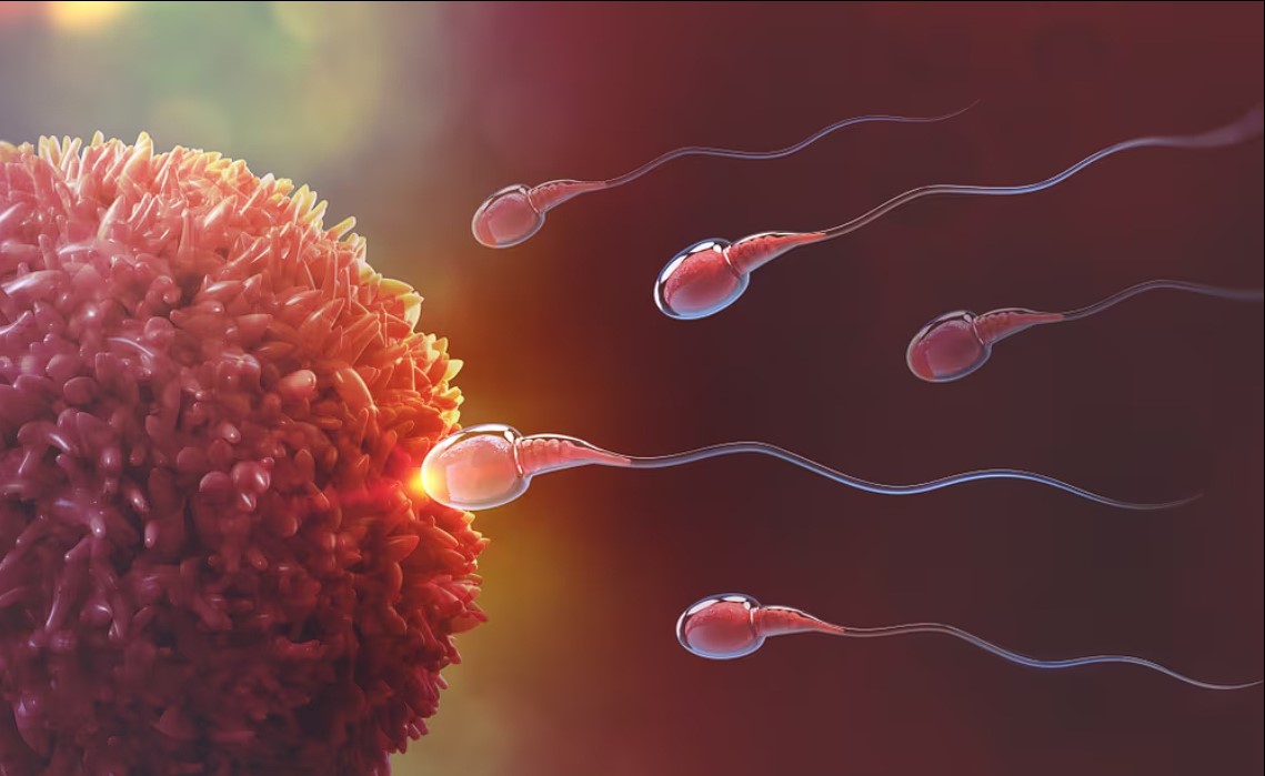
The meeting and joining of two highly specialised cells (gametes), one from a man (the sperm) and one from a woman (the egg), gives rise to life, i.e. the zygote (the fertilised egg).

This amazing journey begins with two cells coming together and creating a kind of cell factory. From this cell, two, then four and so on arise. Up to 8, identical and undifferentiated (they are called totipotent because each of them can give rise to any other cell in the body). Consider that after nine months, this cluster, initially consisting of a few cells, will have increased in size by a factor of 200 billion. Four days after fertilisation, we have just 16 cells (blastomeres) that have not yet implanted into the uterus (pictured). This is the podolocular state, called the morula because of its mulberry-like shape. On day 14, the implantation of what is now called a blastula, consisting of 100-150 cells, is prepared.
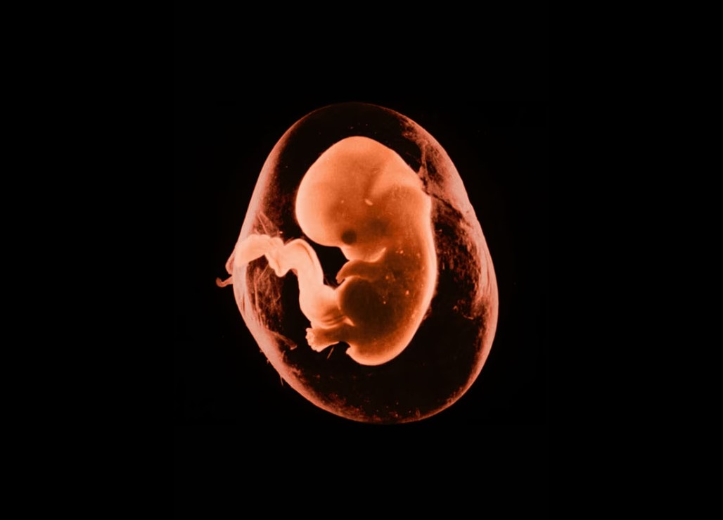
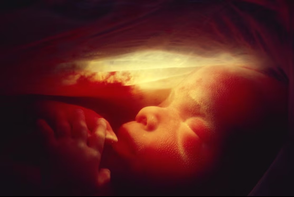
After six weeks, the embryo floats in amniotic fluid, which protects it. It attaches itself to the placenta through the umbilical cord: from here it receives the nutrients and oxygen it needs to develop and carbon dioxide and metabolic waste are removed. The placenta allows small molecules to pass through but not larger ones, such as blood, creating the necessary protection. If, for example, the baby's blood composition is incompatible with that of the mother, an immune response may begin to expel the 'foreign body'. At this stage, the embryo is 13 to 22 millimetres long. The fingers and toes begin to form and separate.
In the eighth week of pregnancy, the body and limbs are developing: even the eyes are already clearly visible. This date marks the watershed between the foetal and fetal phases: at this stage the foetus is able to respond to stimulation around the mouth by contracting the neck muscles and slowly turning the head. This means that the muscles contain nerve fibres: the neurological system has begun to function. At this stage, the differentiation of cells into different organs (organogenesis) is much more important than growth in centimetres: a tiny creature 23 millimetres long and weighing 1 gramme floats in the mother's belly.

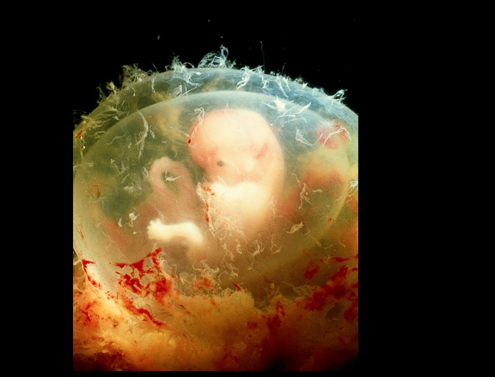
Clinging to the umbilical cord, the foetus grows (6 centimetres long and about 20 grams, between the 12th and 15th weeks) and begins to move its arms and legs in the placenta, which is already fully developed. The eyes form, but the eyelids remain closed. Inside the womb, the foetus is surrounded by complete darkness, although it is probably able to perceive changes in external brightness. Indeed, experiments have shown that directing a beam of bright light onto the mother's abdomen causes the foetus to tremble and its heart rate to increase at 15 beats per minute.
Between the end of the first trimester and the beginning of the second trimester, the foetus cannot yet survive on its own, but it already has developed systems and functions.
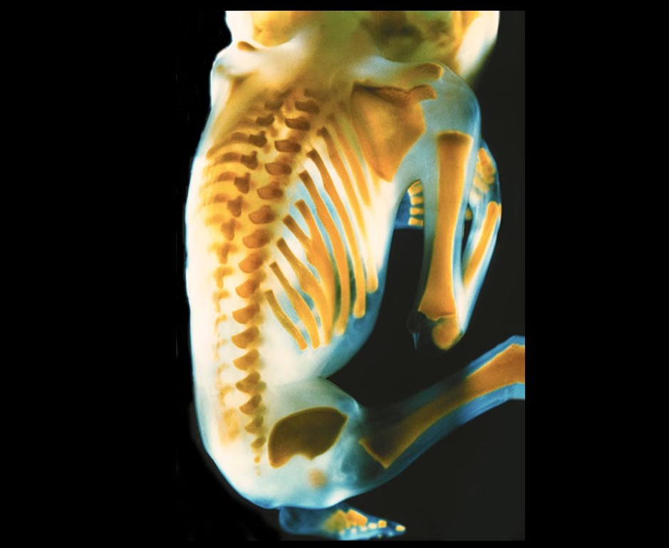
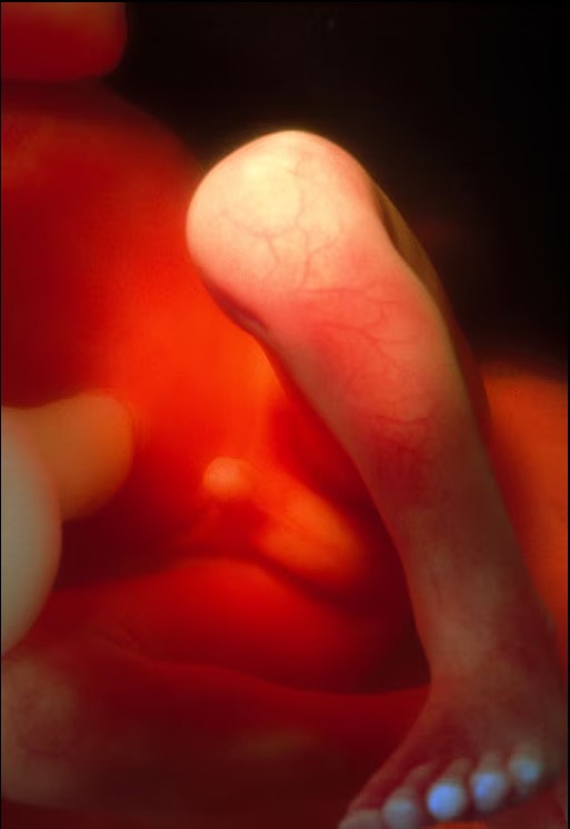
Teeth form inside the gums, nails grow, ribs and vertebrae begin the ossification process by which cartilage turns into bone. In the picture, the hardest bones are the yellowest and most defined bones that appear to be separated from each other. In fact, there is a connection, but it is still cartilaginous and not visible. At this stage of development, the foetus weighs 200 grams.
This is how life begins, halfway through...

The foetus in the nineteenth week of pregnancy is covered with scattered fluff, which will disappear before birth. In reality, some babies are much hairier at birth than others. This is because in the sixth month of pregnancy the hair follicles develop, from which hair is produced in the seventh month. The life cycle of this first hair ends at the same time, and hair falls out all at once: if this happens while still in the womb, the baby will be bald at birth, otherwise his or her hair will be relatively thick. However, these differences are dictated by genetic predisposition.
The face of an embryo is formed very early, as early as 25 days after fertilisation, when three outgrowths, the "branchial arches", emerge from an embryo no bigger than a bean. While in fish these turn into gills, in mammals these three outgrowths merge to form a forehead, face and throat. It is only after a further 6-7 weeks that the face takes on a human appearance: before the last trimester of pregnancy, it finally acquires the characteristic features of an individual. At 20 weeks, the fetus is about 19 centimetres long and weighs 500 grams: we are about halfway there...

From the sixth month onwards (pictured with the fetal ear in the twentieth week), the unborn baby responds to the sounds around it: the regular flow of maternal blood, the steady beat of the heart and the mother's voice lull it through the entire gestation period. But that's not all. If loud sounds come from outside, the baby reacts by speeding up its heartbeat and moving its legs and arms. The foetus is also sensitive to music: nothing soothes it more than its mother singing and listening to classical music such as Mozart or Vivaldi.
As early as the fourth month, the sex of the unborn child becomes apparent: the external genitalia are clearly visible on ultrasound. By 22 weeks, the foetus weighs 650 grams and its length from head to coccyx is 21 centimetres. At this stage of pregnancy, fetal movements are easily perceived by the mother. In addition, the baby is perfecting a technique that will lead it to suckle mother's milk at birth: it unfolds its thumb and begins to suckle it. Later, near the end of pregnancy, the foetus moves its mouth regularly every 10-20 seconds, even during deep sleep: learning continues...

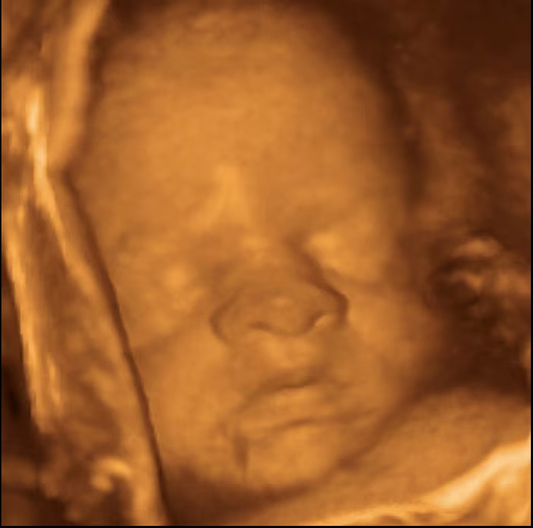
Ultrasound is an ultrasound-based diagnostic method used to visualise the inside of the human body and, in the case of pregnancy, the inside of the uterus. A two-dimensional ultrasound allows you to see the viscera as if in cross-section. The new ultrasound, on the other hand, gives a three-dimensional view of what is going on inside the mother's belly. The photo shows the fetus at twenty-seventh week and already ready to give birth, seen with an MRI. After the twenty-eighth week, the baby is capable of going into preterm labour.
An extraordinary view into the womb is given by a 4D ultrasound, which shows three-dimensional images in motion and in real time: you can see the foetus moving its little hands, sucking its finger or playing with the umbilical cord. The best view is possible after the 25th week, when the foetus reaches of medium size.
Life is an amazing journey, from the moment of conception to the kind of people we become. The wonder of life is in the most ordinary, tiny cells that grow into complex organisms, and in the spark of consciousness that emerges as we develop. We can embrace the wonder of this journey by exploring the world around us, being open to new experiences, learning and growth.
Life is full of surprises, and some of the most magical moments happen when we least expect them. So take the road less traveled, try new things and never stop exploring new things. Let us enjoy every moment and create life and destroy it. Remember, every next day could be your last on this planet. Be daring, live and be yourself.
 and then